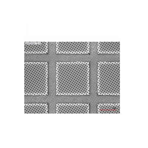
Graphene oxide (GO) provides a thinner support film of higher mechanical strength, electrical and thermal conductivity compared to support films made from other materials. GO support films are available on lacey carbon grids and Quantifoils.
300 mesh copper lacey grids available with 4 thicknesses of CVD graphene. Graphene coverage of the TEM grid is better than 75%. Supplied as a pack of 6 in rotary storage grid box.
Graphene oxide membranes are robust and almost transparent under an electron beam, making them ideal supports for the analysis of a wide range of samples by transmission electron microscopy (TEM). As produced, they are hydrophilic but this can readily be tuned as required. The graphene oxide membranes give a crystalline diffraction pattern similar to graphene and this structure acts as a convenient calibration for electron diffraction and high resolution TEM. Their ultra-low contrast makes them particularly useful for imaging small nanoparticles or nanowires whose structure is not readily resolvable on conventional carbon supports [1]. They can also be used for imaging and analysis of polymeric, macromolecular and biological samples without the need for heavy metal staining [2].
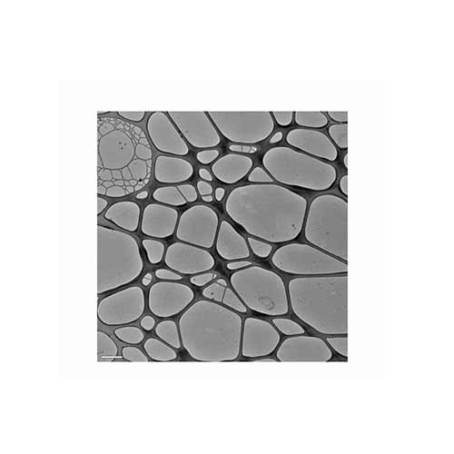
Graphene oxide support grid morphology: the graphene oxide membranes are deposited on lacey carbon supports. Monolayer graphene oxide membranes can span the holes in the lacey carbon. The graphene oxide sheets are distributed randomly across the EM grids such that some holes in the lacey carbon support are uncovered, some covered by a single monolayer of graphene oxide, and some by two or more layers of graphene oxide. On average, roughly half to three quarters of the lacey carbon holes are spanned by graphene oxide and of those roughly half are monolayer.
Structure and composition of graphene oxide: graphene oxide is composed of a graphene like sheet randomly decorated with oxygen functional groups such as epoxy and hydroxyl. Due to its graphene-like carbon backbone, graphene oxide is strong enough to span the holes in a lacey carbon support even when only a monolayer thick. The graphene oxide is synthesized from graphite powder using a modified Hummers’ method.
Use of graphene oxide grids for TEM imaging and diffraction: The ferritin was deposited from aqueous solution by placing a drop on the graphene oxide coated TEM grid and allowing it to dry. As produced, the graphene oxide is hydrophilic. However, this can readily be changed by heating in air if a hydrophobic surface is required. The crystalline graphene backbone gives a characteristic diffraction pattern which can be used as a convenient calibration for the analysis of other samples either by high resolution TEM or electron diffraction.
Identifying the graphene oxide membranes: one monolayer thick the graphene oxide is almost transparent under the electron beam and so is often only apparent through wrinkles or creases in the film. Overlapping sheets are also readily visible. The most distinctive way of identifying monolayer thick regions is from the diffraction pattern: a single layer has a hexagonal diffraction pattern similar to graphene whilst multiple layers are usually in different orientations so that multiple rotationally misaligned hexagonal patterns are observed in the diffraction pattern. However, this can also be caused by creases in a single sheet so some care must be taken with this interpretation.
.

 Products
 SERVICES
 Software